As we explained in our previous article, in recent years we have witnessed a true digital revolution in all areas of life.
When we speak of odontology 4.0, any equipment used in the diagnosis process, the planning or the treatment itself involves the use of digital technology.
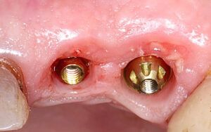
It is more and more common to have some method of taking digital impressions in the clinic, as it helps improve the patient’s experience and evidently facilitates the transition toward a quicker and more stable workflow, notably improving communication with the patient.
To start out on this path to digitisation we recommended the Medit range of intraoral scanners. The company is a pioneer in facilitating access to digital odontology with high-performance scanners at very accessible prices.
And just as important as having an instrument that suits your needs is having the knowledge to use it correctly, to get the most out of it and thus prevent errors when taking impressions that will affect the treatment further down the line.
For this reason, Medit recommends following its simple 7-STEP protocol for scanning the maxilla, the mandible and the occlusion.
Shall we start?
Easily scan the maxilla and the mandible in the same way:
Step 1: Create the basis for the principal scan by scanning the occlusal surface from last molar to the front teeth.

- Scan the lingual side of the front teeth, rotate the tip to scan the incisors, and tilt it again to scan the labial side.
- Complete the front teeth and continue scanning the occlusal surface until you reach the opposite molars.

How do we scan the occlusal surface of the mandible?
Step 2: When you reach the opposite molar, tilt the tip toward the lingual side so that the lingual side of the teeth, the gums of the lingual part and the occlusal surface appear as 1:1:1, and scan the lingual side from the opposite molar to the molar where you started scanning.

- If you tilt the scanner too much to the lingual side, the system can get lost due to the lack of characteristic areas.
Step 3: When you finish with lingual side, tilt the tip toward the vestibular side so that the vestibular side of the teeth, the vestibular gums and the occlusal surface appear as 1:1:1, and scan the vestibular side to the opposite molar.

Maxillary vestibular side Scanning process |Scanning process of the vestibular side of the mandible
- The more scan data there is of the gums, the easier it will be for the software to align them later.
Once the scanner has finished the maxilla and the mandible, start with the occlusion. But first, check the following:
·Seat the patient in a vertical position, with the occlusal plane parallel to the ground.
·Check whether the scanner data of the maxilla and mandible have unnecessary soft tissues. If so, eliminate them with the cutting tools.

·Check whether there are gaps in the scan data of the maxilla and the mandible. If so, conduct further scans.
Step 4: Place the tip of scanner in the middle of the deepest posterior area of the maxilla and the mandible. Start the occlusion scan using the roller method: rotate the tip up and down until you have scanned 3 to 4 teeth from the maxilla and the mandible, respectively.
- Take the scan data of the similar posterior teeth for the occlusion of both quadrants.


Step 6: If the system acquires sufficient 3D images, the alignment will be carried out automatically.

Step 7: The initial alignment is completed for occlusion.
- Using the process of automatic or manual alignment, the maxilla and the mandible are placed in the initial position to facilitate alignment.

+ Bonus Track:
· Medit has a display mode called “reliability map”. To know whether the scan is reliable, it must be displayed in green, as shown in the following image.
· The scan areas that do not have any metal restorations or edentulate zones are suitable for occlusal scanning. If the area selected has characteristic shapes, it is advantageous for the occlusion process.
If you need more information about how to scan properly and have the new Medit i700, you can start with “Practice mode”, which will help you perfect your intraoral scanning through an exercise with different levels of training (for beginners, competent users and experts).
Finally, if you need personalised consultancy, you can contact our technical team, we are always happy to resolve any doubts you may have.