Description of the case
Esthetic crown lengthening: Female, 19 years of age, with no medical history of interest or harmful habits, presenting excessive gingival exposure upon smiling.
After an intraoral scan and cone-beam computerized tomography, a customized periodontal surgical guide was co-designed with Avinent using the DentalCad (Exocad) software. The polyamide device manufactured by Avinent presented an inner cutting area corresponding to the future cervical gingival margin, and an outer area that indicated the maximum ostectomy to be performed.Esthetic crown lengthening.
Incisions were made with an internal bevel of 1.5 to 2.5 respecting the indications of the surgical guide. Then the device was removed to finish edging the tissues with a diode laser. Then a full-thickness gingival flap was opened, the guide was repositioned, and the osteoplasty and ostectomy were carried out following the outer cutting area (3 mm from the amelocementary line up to the crestal bone). Finally, the papillae were sutured with vertical mattress sutures. No intra- or post-operative complications were reported.
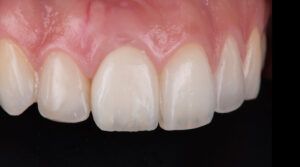
The clinical follow-up at 4 months showed complete tissue stability, as well as the balance and harmony of the smile. The patient’s desired esthetic result had been achieved.

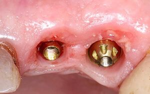
Initial photos of the case Initial photos of the case Initial photos of the case Initial photos of the case


Design of the surgical guide
Planning the height of the final crowns | Image of the final resultDetails of the fit and thickness of the surgical guid


Image of the surgical guide in the mouth Guided incision of the tissues


Outline of the vestibular incisionRemoval of the marginal tissue with a rasp Laser shaping of the tissues


Result of the planned gingival margin Result of the planned gingival margin


Details of the flap detachment Details of the flap detachment Detachment of the full-thickness flap Repositioning of the guide for ostectomy


Details of the ostectomy and osteoplasty Verification of the distance between the alveolar bone and the amelocementary line Repositioning of the flap and suture Verification of the final result with the surgical guide


Healing after 15 days Suture removal


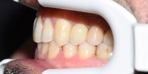
Final outcome of the case Final outcome of the case Final outcome of the case


Final appearance


Design of the surgical guide Planning the height of the final crowns | Digital image of the final result Details of the fit and thickness of the surgical guide Initial photos of the case
Initial photos of the case
Initial photos of the case
Initial photos of the caseImage of the surgical guide in the mouth Guided incision of the tissues Outline of the vestibular incision Removal of the marginal tissue with a rasp Laser shaping of the tissues Result of the planned gingival margin Result of the planned gingival margin Details of the flap detachment Details of the flap detachment Detachment of the full-thickness fla Repositioning of the guide for ostectomy Details of the ostectomy and osteoplasty Verification of the distance between the alveolar bone and the amelocementary line Repositioning of the flap and suture Verification of the final result with the surgical guide Healing after 15 days Suture removal Final outcome of the case
Final outcome of the case
Final outcome of the case
Final outcome of the caseFinal appearance Case comparison before and after