Introduction
Knowledge of the processes involved in osseointegration has enabled good progress in dental implant treatments, with a very positive impact on edentulate patients in particular. It provides an alternative to restorations with conventional complete prostheses and the functional inability that this entails, above all in the mandible.
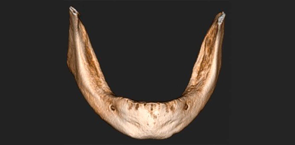
This type of patient usually presents mandibular reabsorption with narrow socket crests as a consequence of complete edentulism for a long period of time. This situation, alongside the existence of anatomical structures such as the dental nerve, can increase the complexity of implant treatments, forcing us to use more complex techniques to guarantee a good result.
The success of these complete fixed restorations with implants is high provided there is good maintenance and control of the periimplant tissues.
Clinical case
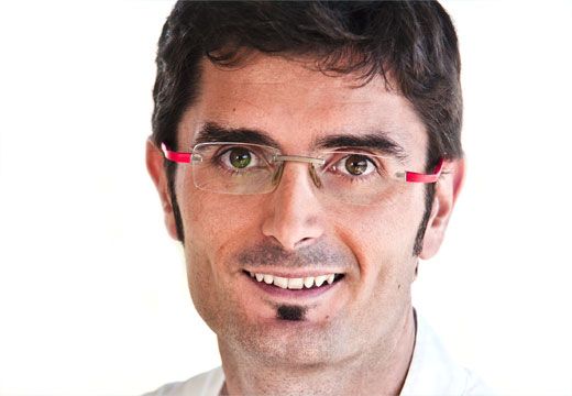
Edentulate patient aged 87 years, comes to the clinic as she cannot tolerate the mobility of the complete lower prosthesis she has had for more than 10 years.

Initial situation of the patient. Initial situation of the patient.
Procedure
After explaining to the patient that the bone reabsorption in the mandible hinders the stability of the prosthesis, we proposed fitting four Avinent Biomimetic Iceberg implants and converting her complete prosthesis (CP) into a fixed implant-supported overdenture.
To do this in a minimally invasive way, it was decided to use guided placement for the implants. Using a functional digital impression of the patient’s CP with Permlastic (Kerr), the STL was printed to make a radiographic splint with gutta-percha markers using the dual scan protocol. The position of the implants was planned with Implant Studio (3Shape) software and a mucosa-supported surgical guide with two anchor pins was made.

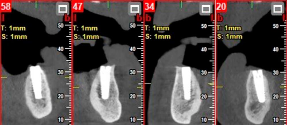
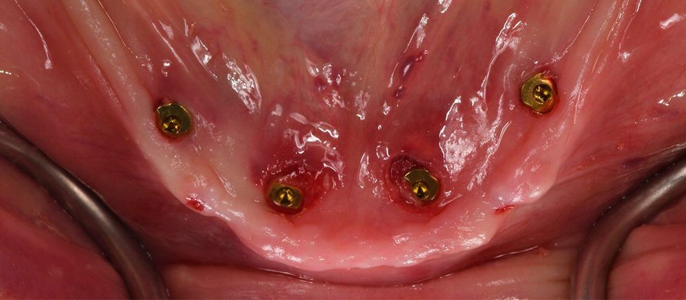
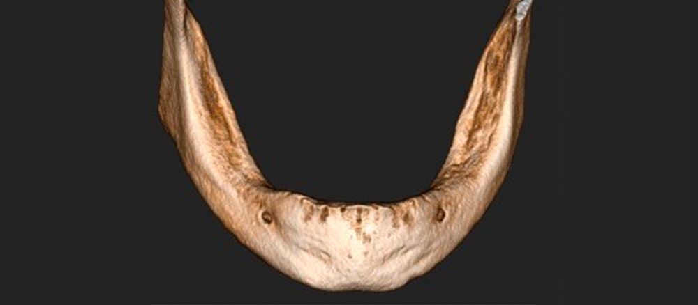
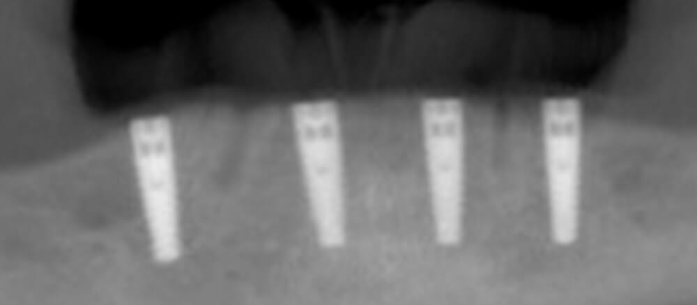
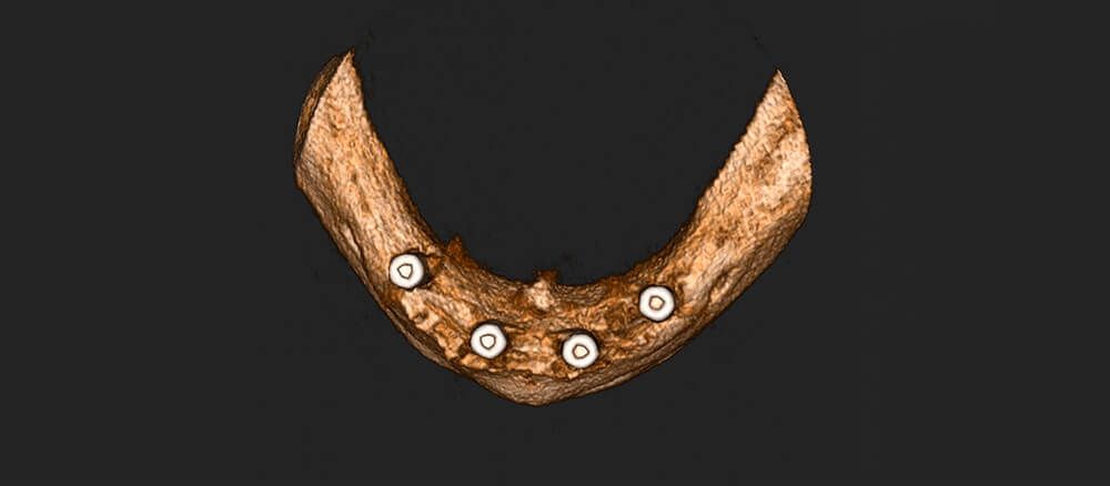
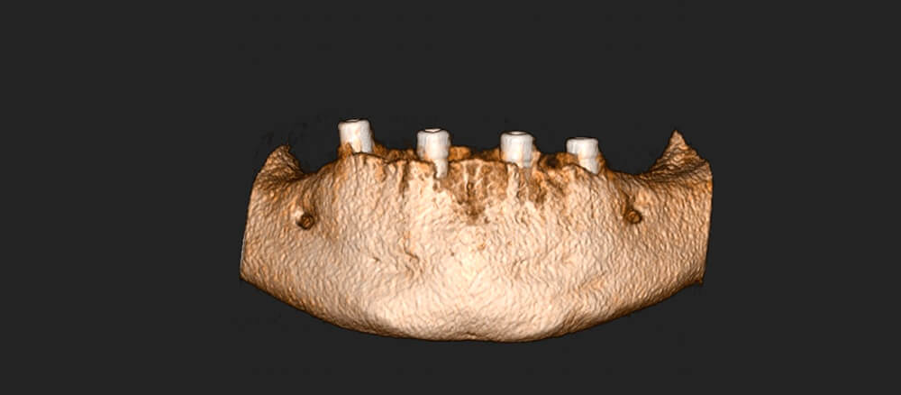
Four Biomimetic Iceberg 3.5 x 11.5 mm implants were placed in the positions 34, 32, 42 and 44. Even though we achieved a torque of 40 N in all of them, we chose to place the healing caps and use the complete prosthesis as the provisional for healing.
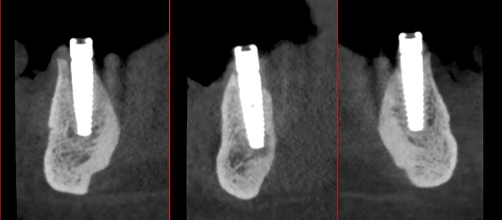
Implants after transmucosal placement X-ray view of the implants in the maxilla X-ray view of the implants in the maxilla


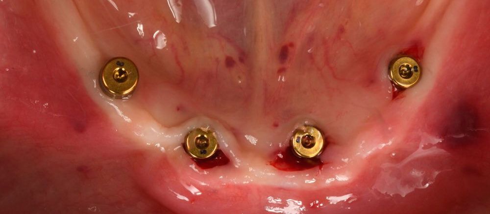
After a period of 3 months, in a second stage surgery, healing abutments were placed and the patient’s complete prosthesis was temporarily rebased.
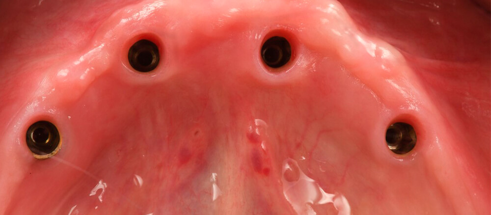
Placement of the healing abutments in a 2nd stage surgery after 3 months Placement of the healing abutments in a 2nd stage surgery after 3 months

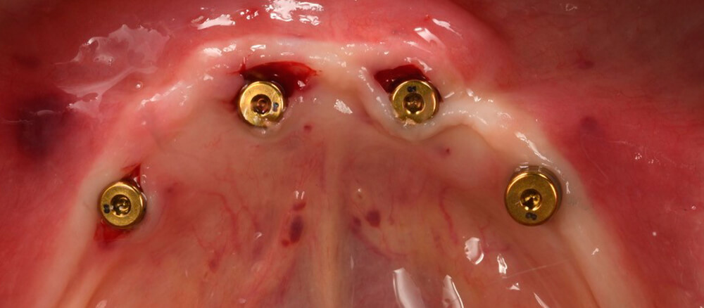
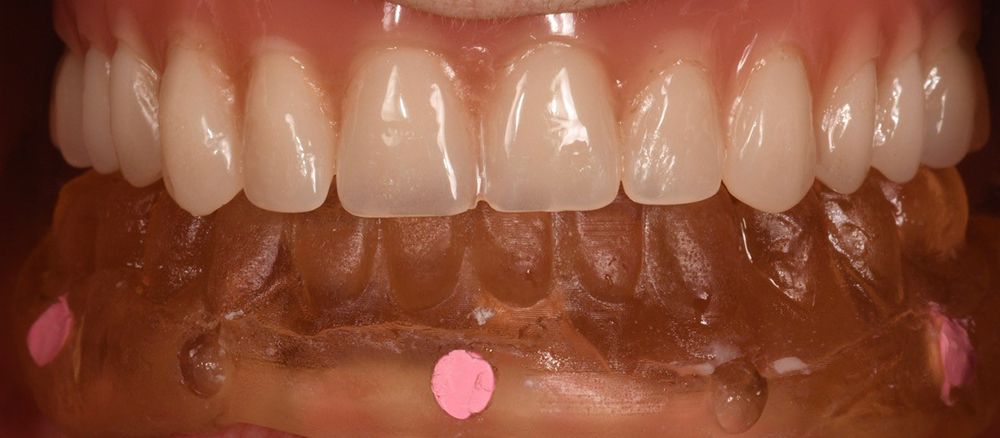
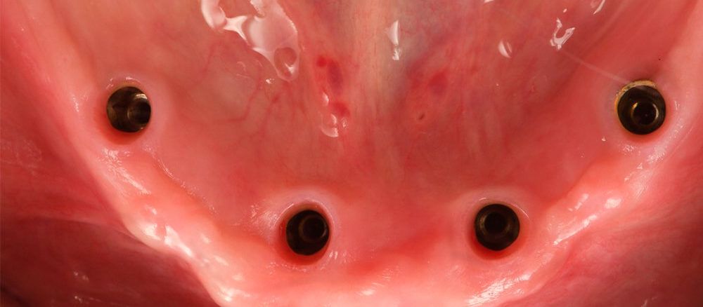
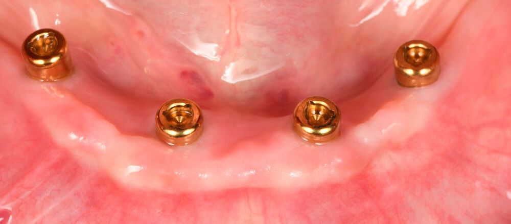
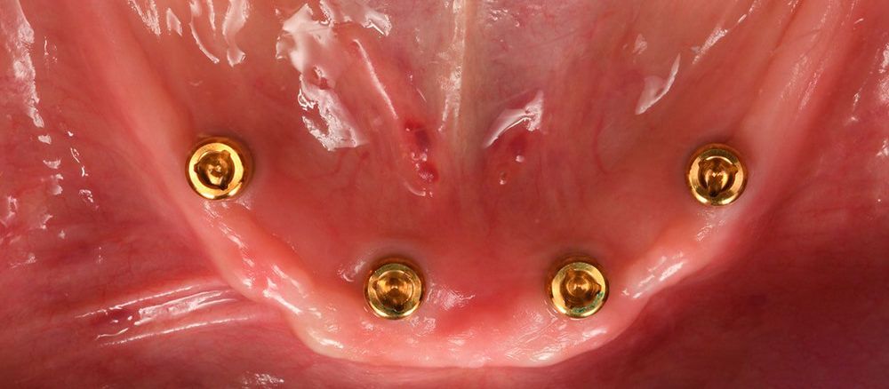
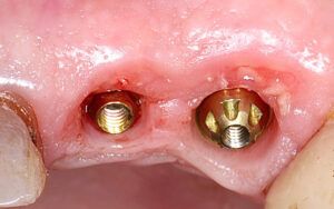
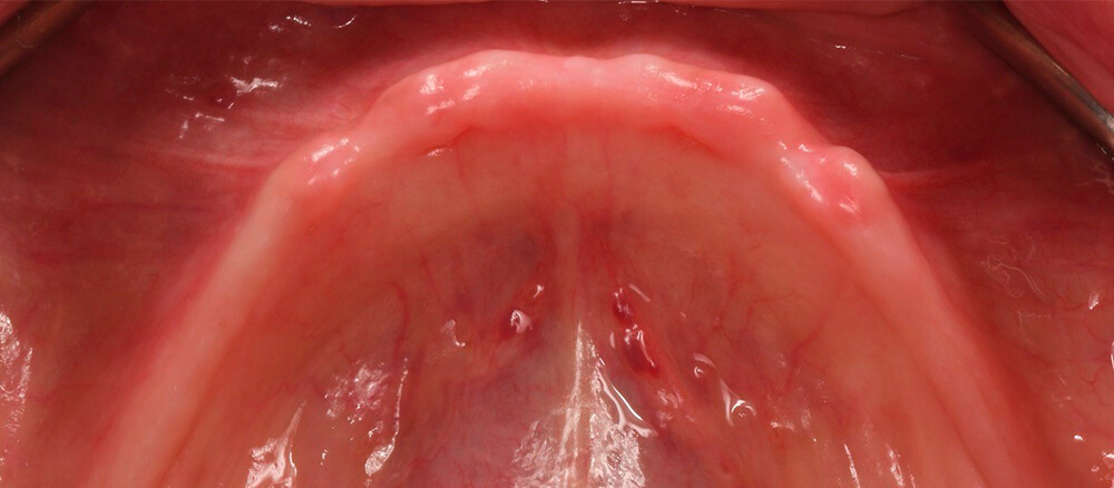
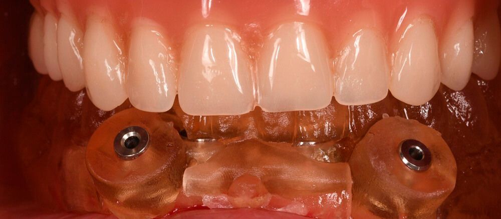
The healing stage did not result in any complications and once complete we fitted the 4 Locator abutments to convert the patient’s CP into a removable implant-supported overdenture. The implants were distributed according to the patient’s own prosthesis for good retention.
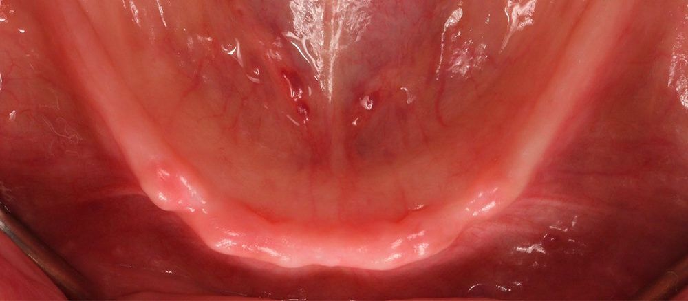
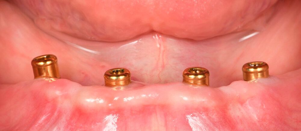
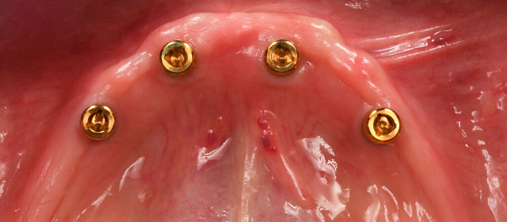
Scan of the Locator abutments in the mouth Scan of the Locator abutments in the mouth Details of the condition of the tissues
Images of the tissue health with the overdenture abutments screwed into the implants Images of the tissue health with the overdenture abutments screwed into the implants

Conclusion
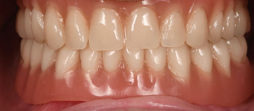
Implant treatments on narrow socket crests, a consequence of edentulism over a long period, are usually complex. In this case, with prior digital planning and a minimally invasive treatment, we were able to provide a solution to the patient’s problem with her complete lower prosthesis.
The use of Biomimetic Iceberg implants enabled us to provide stability in the soft tissues, producing a peripheral seal and achieving a good clinical result, preserving a ridge of keratinized gum for correct maintenance of the patient’s implants in the medium and long term.


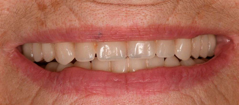
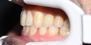
Initial situation of the patient Initial situation of the patient Duplicate of the patient’s CP with gutta-percha markers Placement of the surgical guide in the mouth Implantes postcolocación transmucosa X-ray view of the implants in the maxilla Placement of the healing abutments in a 2nd stage surgery after 3 months Placement of the healing abutments in a 2nd stage surgery after 3 months Details of the condition of the tissues Scan of the Locator abutments in the mouth Scan of the Locator abutments in the mouth Images of the tissue health with the overdenture abutments screwed into the implants Images of the tissue health with the overdenture abutments screwed into the implants Image of the final removable prosthesis Images of the tissue health with the overdenture abutments screwed into the implants