
Introduction
Dental impaction refers to the anomaly in which a tooth does not erupt after reaching full anatomic development.
After the wisdom teeth, the canine is the tooth that is most often impacted, with a prevalence of 0.98-2.27% of the population, and a higher frequency in women (1.17%) than men (0.51%). In addition, it is more frequent in the maxilla than in mandible and cases of retention are found more to the palatine than vestibular. Bilateral impactions occur in 8% of cases.
The etiology of retained teeth has multiple factors. It may be due to local causes such as a lack of space and alterations in tooth eruption, ankylosis, trauma to adjacent teeth or root dilaceration among others.
Systemic causes include a genetic predisposition, cleft lip, vitamin A and D deficiency and syndromes such as Crouzon and Down.
Clinical case
Reason for consultation and diagnosis
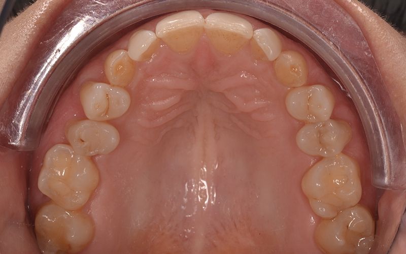
Patient aged 25 years, without no notable medical history, comes to the clinic due to bilateral impaction of the maxillary canines with prolonged retention of the deciduous canines.
In addition, a bilateral crossbite and a dental and skeletal class III were also noted. No alterations were presented at periodontal level.

Clinical examination
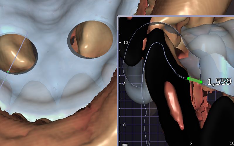
Clinical and radiological (CBCT) examinations were carried out to locate the canines in three dimensions, assess their status and relationship with the roots of the neighboring teeth, and determine the possibility of orthodontic and surgical treatment.
Treatment options
Depending on the type of retention (palatine or vestibular), the three-dimensional position of the canine, its relationship with the adjacent teeth and the patient’s age, we recommend fenestration and traction of the canine or extraction.
In this case it was decided to extract the canines using surgical guide designed for this purpose.
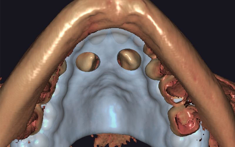
Planning of the case
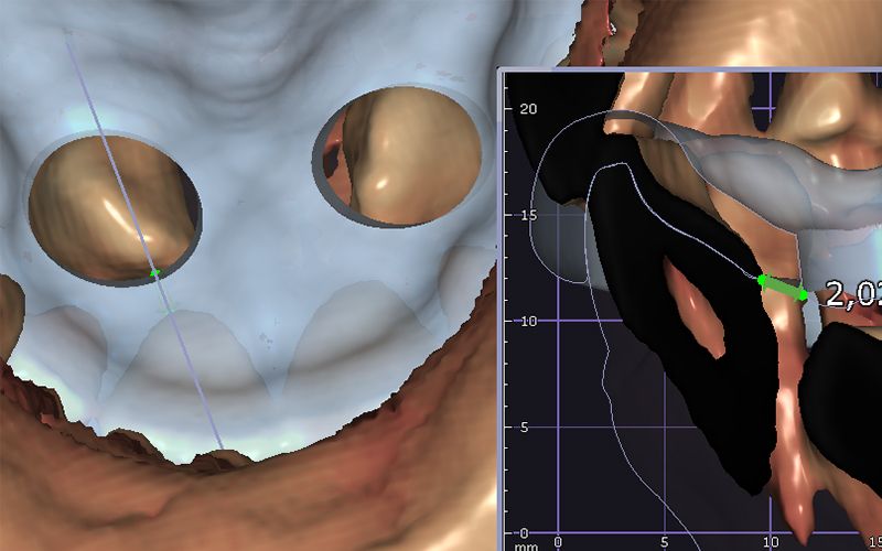
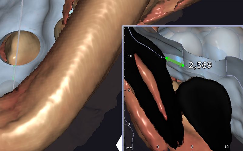
A CBCT and an stl. file of the patient’s soft tissues and teeth were obtained with an intraoral scanner. The two files were superimposed and the surgical guide was designed using the DentalCAD Rijeka dental design software (Exocad, Germany) at the Avinent CadCam (Avinent Implant system) milling center.
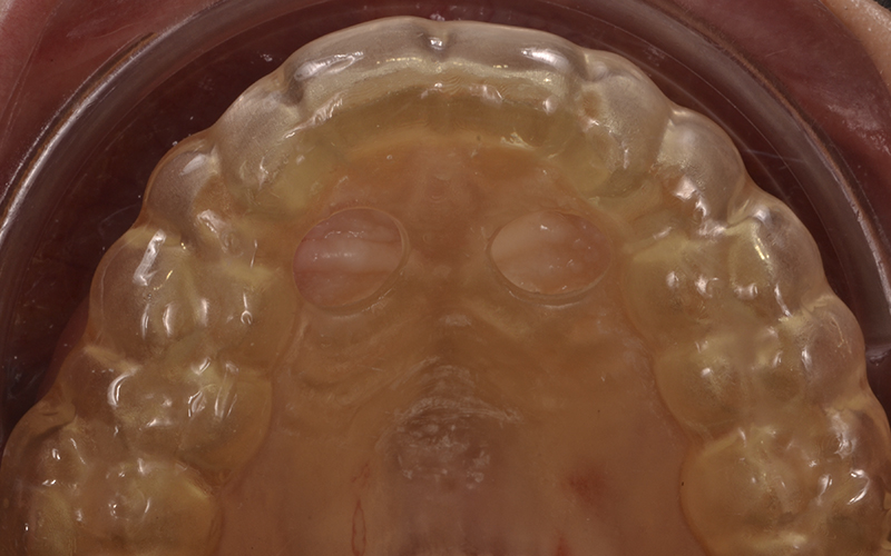
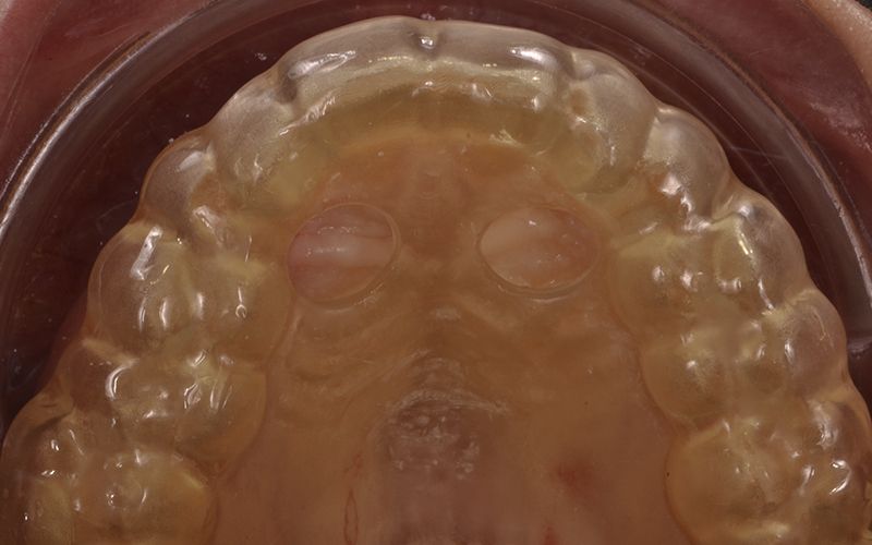
The design must take into account the depth of the canine and its 3D position in relation to the adjacent teeth, to be able to design the window and delimit its opening. Ideally the guide window must delimit and expose the crown of the impacted canine.

Details of the dental design software Details of the dental design software Digital design of the surgical guide


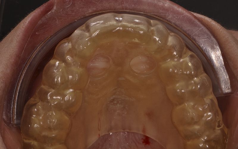
To manufacture of the guide, we used the CAM Formlabs PreForm software (Formlabs, USA) and printed it with the Formlabs Form3B 3D printer (Formlabs, USA) in Dental SG Resin biocompatible resin for surgical guides (Formlabs, USA) that can be sterilized in an autoclave, using Avinent CadCam (Avinent Implant system).
Surgical technique
Under infiltration anesthesia, we verified the fit of the guide.
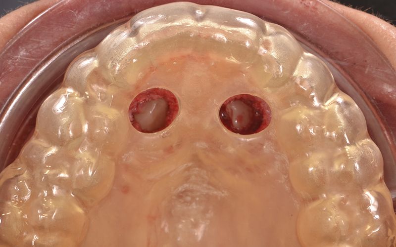
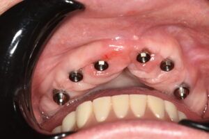
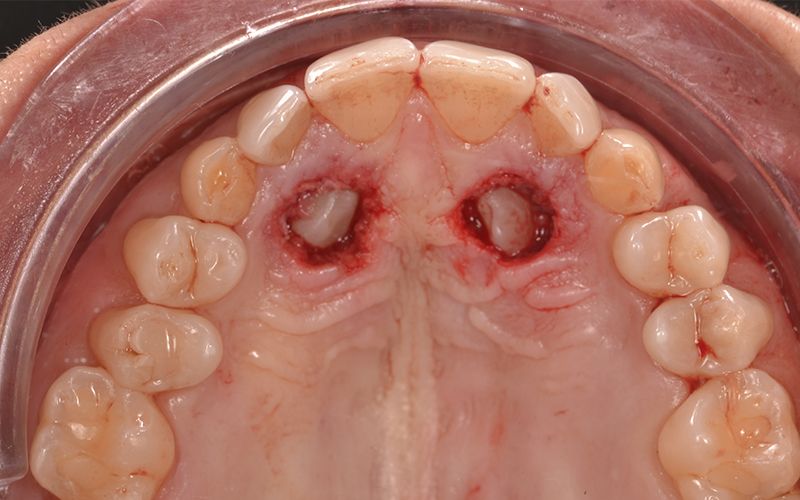
Then the soft tissue was eliminated from the mid-palate and the ostectomy was performed to expose the crown of the tooth.

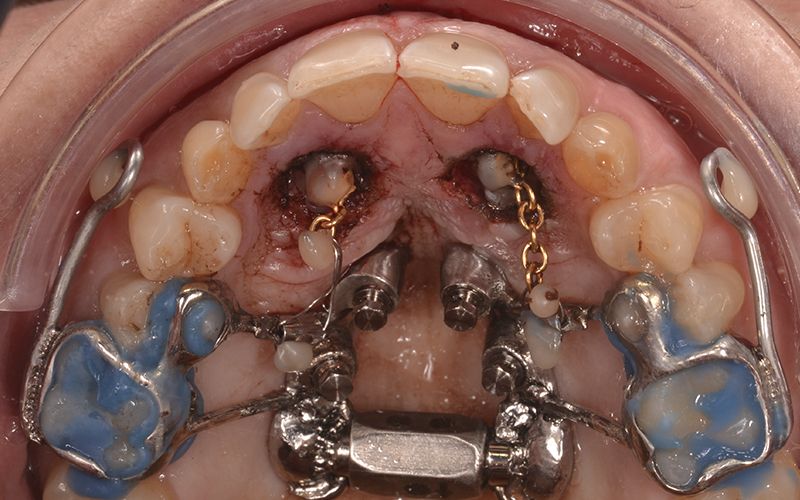
Exposure of the bilateral canines through elimination of soft tissue Exposure of the bilateral canines through elimination of soft tissue

A dental circuit breaker was used as an anchor point with a gold chain affixed to the surface of the tooth by acid-etching, adhesive and compound resins. For traction, a titanium-molybdenum-steel (TMA) rod-type spring was used.

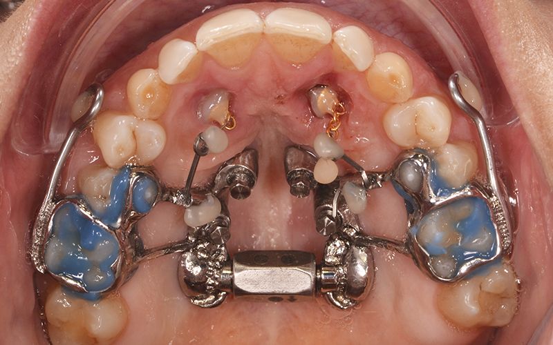
The aim of the treatment was to apply light intermittent forces that would act at the center of resistance of the tooth for:
- Movements toward the mid-line and distal, to disimpact the crown of the canine, using a TMA rod-type spring connected to a micro-screw.
- Movements of traction toward the space the dental arch should occupy.
- Movements of alignment and levelling.
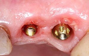
The case is currently in the last stage of treatment, and no post-operative complications of any type have been reported during the treatment period.
Palatine view for reference where the deciduous canines had not erupted Details of the dental design software Details of the dental design software Digital design of the surgical guide Adjustment of the surgical guide on the palate Exposure of the bilateral canines through elimination of soft tissue Exposure of the bilateral canines through elimination of soft tissue Adhesion between the traction component for the canines and placement of the traction medium Image one month after starting treatment