Introduction
One of the situations we face in our daily practice is limited interdental mesio-distal space that hinders the placement of standard metric implants.
To solve this problem and avoid having to resort to dental-supported fixed prostheses or removable prostheses, nowadays we have the possibility of using reduced diameter implants that, as we will see below, are an excellent tool for cases in which the distribution of space is our main difficulty.
Clinical case
Female patient aged 16 years consults the clinic for agenesis of the upper lateral incisors 12 and 22. The patient had no prior medical history of interest and the x-ray and clinical examination revealed excellent oral health. We referred her to the orthodontist who, after the pertinent study, started treatment with braces to try to gain maximum space between the central and canine teeth with the aim of placing two implants in future.
After two years of orthodontic treatment, the patient’s the situation was reevaluated and it was decided to wait approximately one more year before placing the implants. The reason was to ensure the full development of the patient’s maxillary bone.

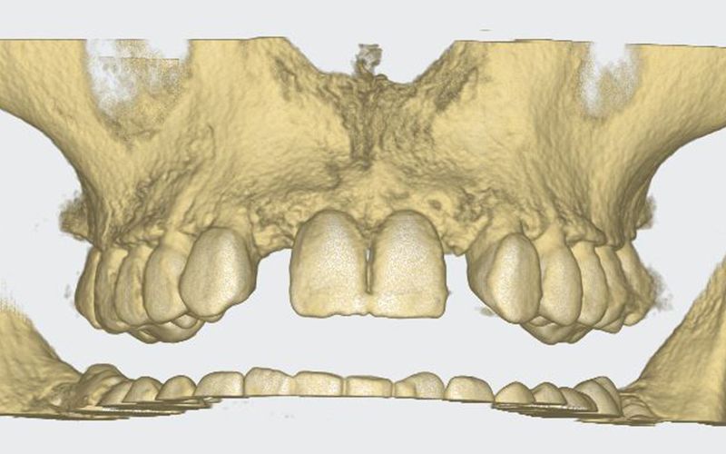
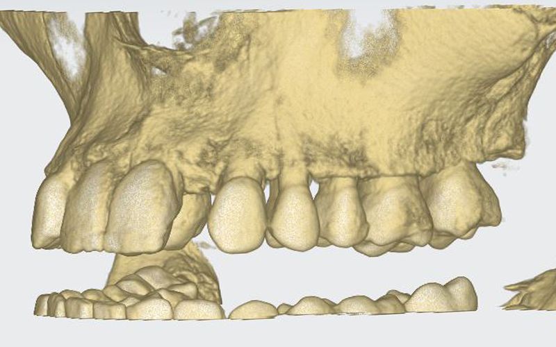
Patient’s initial CT scan after orthodontic treatment Patient’s initial CT scan after orthodontic treatment 
Patient’s initial CT scan after orthodontic treatment


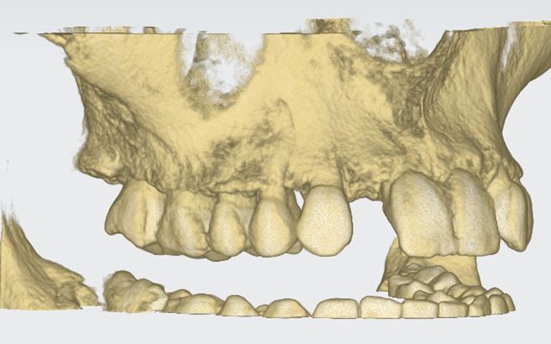
STL images for planning STL images for planning STL images for planning


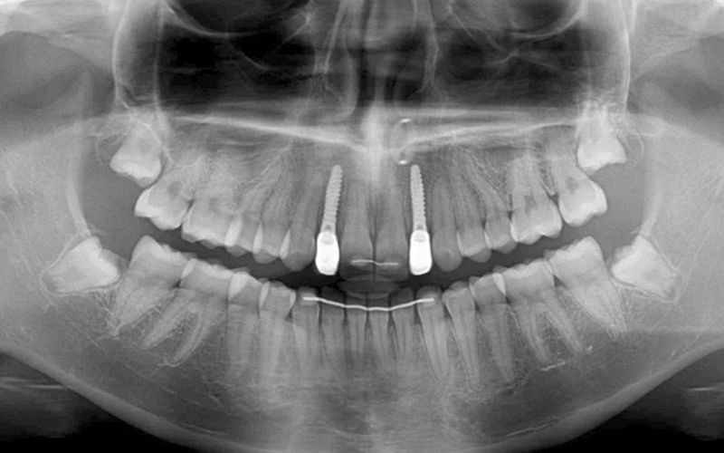
After this time a CBCT was taken of the maxilla to start planning the surgery for the placement of two implants in positions 12 and 22. During the analysis of the x-ray images, as the measurements indicate, it was observed that the mesio-distal space between 11-13 and 21-23 was limited, which would impede the placement of two standard implants with a diameter of 3.3 mm.
Given the circumstances and seeking optimum resolution of the case, it was decided to use Biomimetic Pearl implants with a reduced diameter of 2.8 mm by Avinent Implant System. These mini-implants have a prosthetic line for single restorations and, within their wide range, we found them to be the best choice for the case requirements, as they allow the distribution of space to be perfectly maintained and the biology of the restoration to be respected.
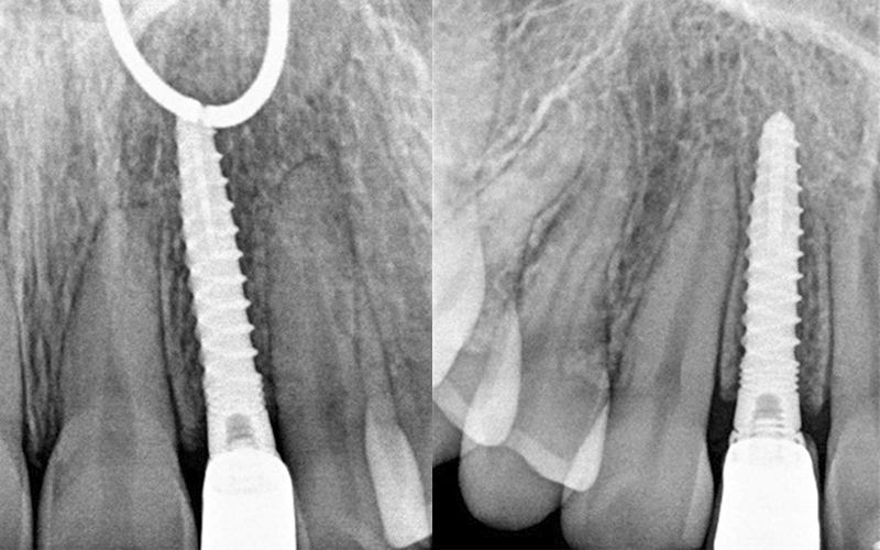
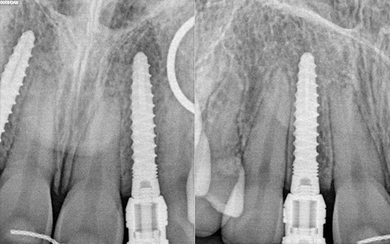
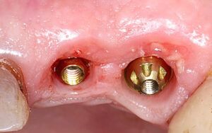
The surgery was carried out for the simultaneous placement of the 2.8 x 13 mm Pearl implants in positions 12 and 22 following the protocol indicated by the company. They were inserted successfully despite the fact that the anatomical conditions were not ideal, with a torque of 30 Ncm achieved in both.

During the osseointegration period of the implants and up to the start of the prosthetic stage, the patient used the provisional prosthesis she had worn since the completion of the orthodontic treatment. The prosthesis manufactured by the laboratory was a Vacupress splint with two resin teeth to cover the absences of 12 and 22.Post placement x-ray of the Pearl implants.
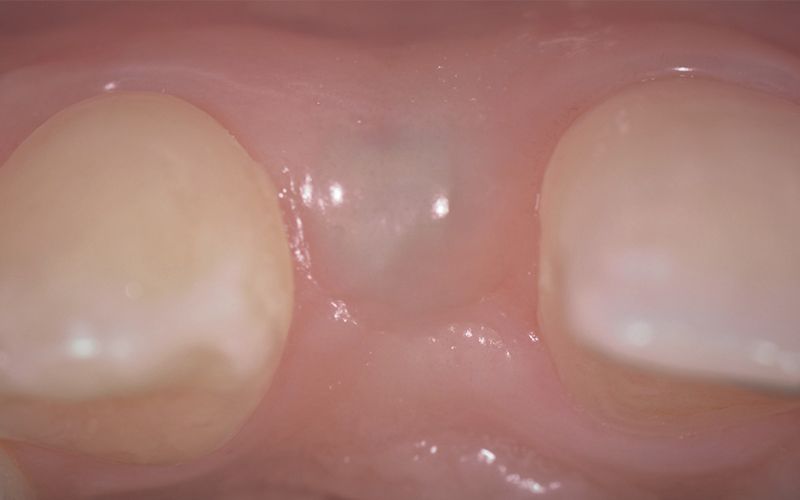
Images of the osseointegration period of the implants and healing of the tissues Images of the osseointegration period of the implants and healing of the tissues Images of the osseointegration period of the implants and healing of the tissues Images of the osseointegration period of the implants and healing of the tissues


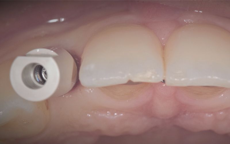
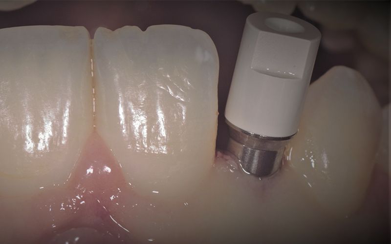
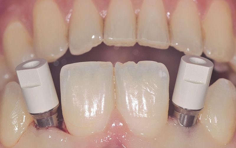
After three months of osseointegration, the second surgery performed on both implants and the digital impressions were taken with the TRIOS (3Shape) intraoral scanner.
Details of the digital impression with the scan abutments placed in the mouth Details of the digital impression with the scan abutments placed in the mouth Details of the digital impression with the scan abutments placed in the mouth


The impressions were sent to a reliable prosthetics laboratory to make provisionals while they worked on the CAD-CAM design and manufacture of the definitive zirconium crowns.
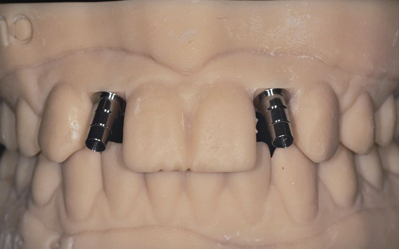
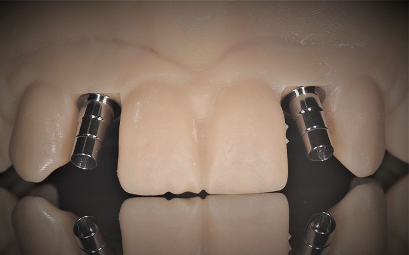
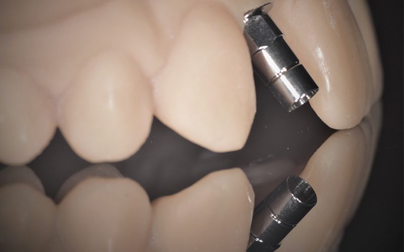
The milled monolithic zirconium crowns with cut-back were cemented on two titanium bases previously cut to correct the insertion axis of the implants.
Details of the laboratory work with titanium bases on a digital model printed in 3D Details of the laboratory work with titanium bases on a digital model printed in 3D Details of the laboratory work with titanium bases on a digital model printed in 3D


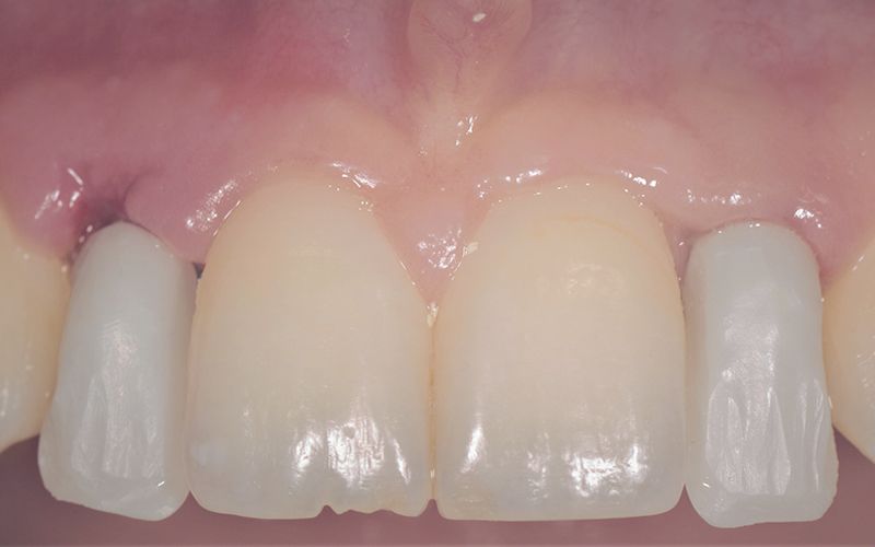
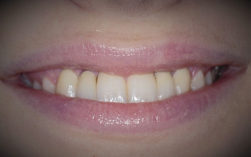
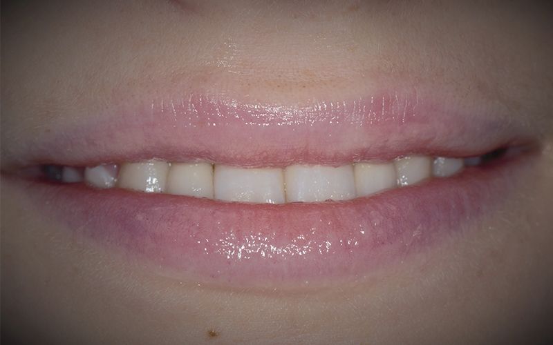
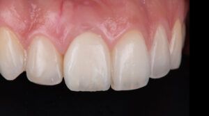
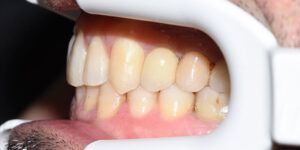
After a few days, the pertinent x-ray and clinical controls were carried out and the two crowns were definitively cemented, completing the restoration of the agenesis of the two lateral incisors 12 and 22.
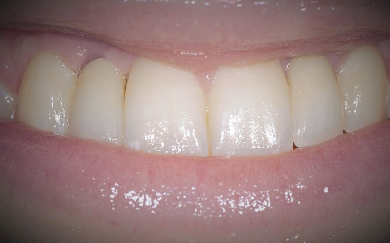
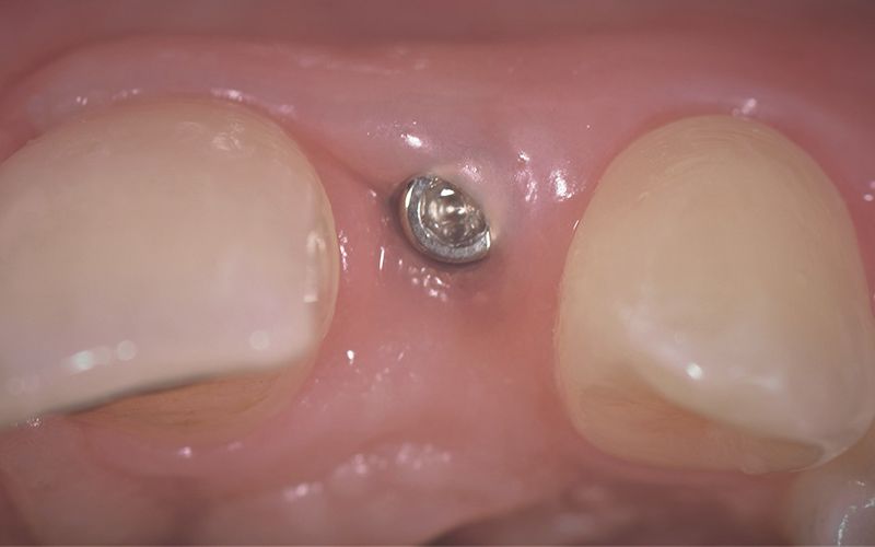
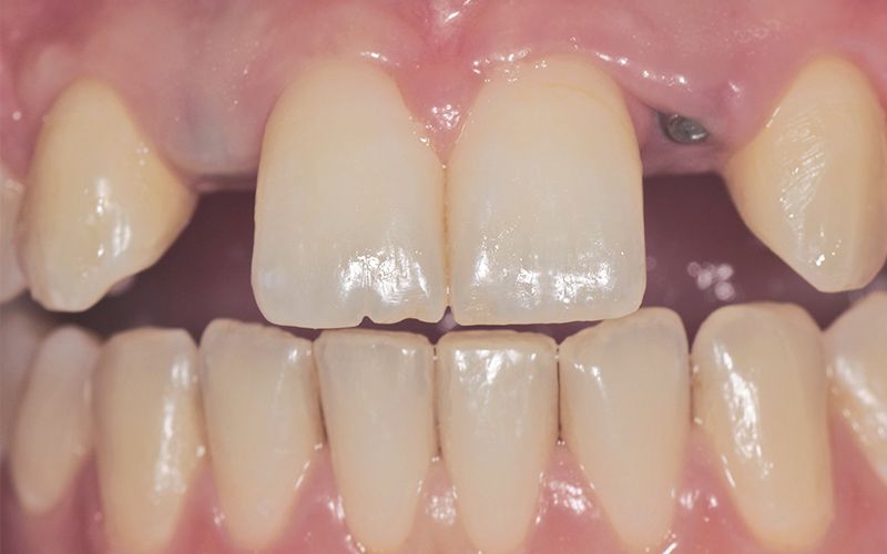
Definitive Zr crowns post-load
Definitive Zr crowns post-load
Definitive Zr crowns post-load
Definitive Zr crowns post-load


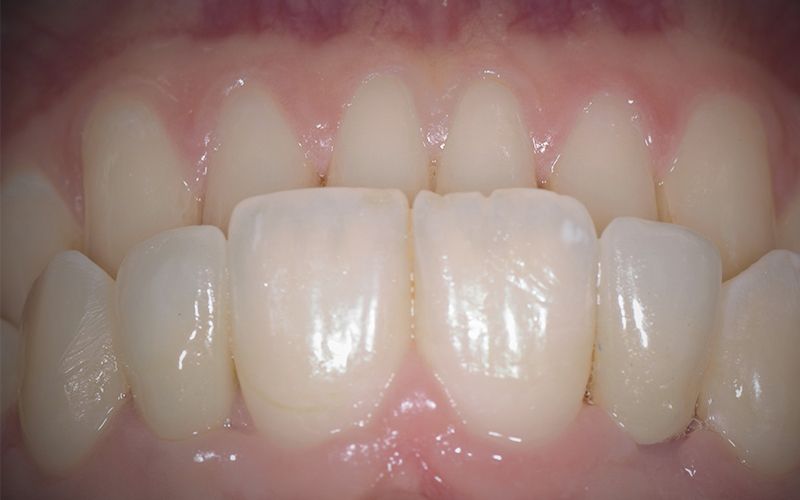
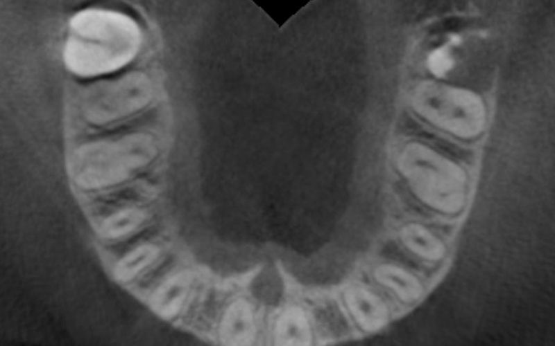
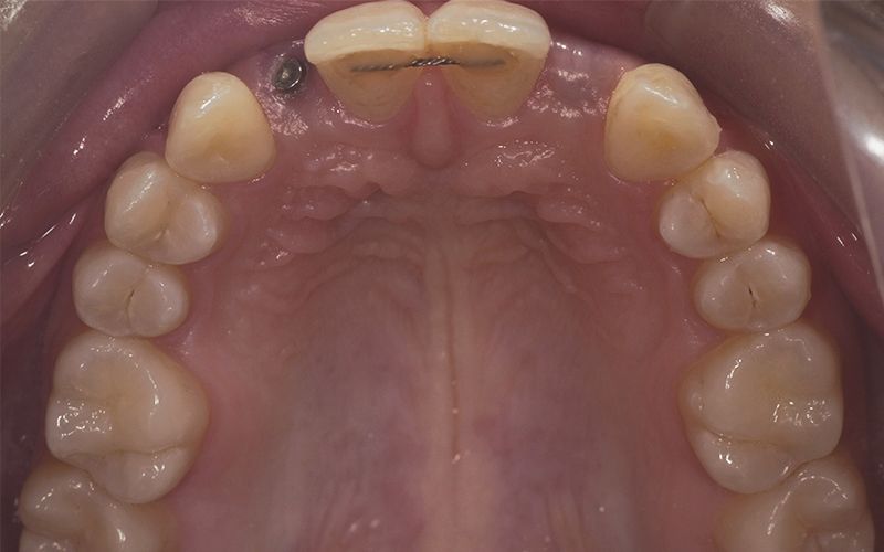
X-ray images of the revision at 6 months post-placement of the implants and 2 months after prosthetic load X-ray images of the revision at 6 months post-placement of the implants and 2 months after prosthetic load


The Biomimetic Pearl system enables minimally invasive treatment of single cases with limited space resulting in a final restoration that meets the patient’s functional and aesthetic expectations.
Currently the good clinical behaviour of mini-implants, with diameters under 3 mm, means they can be used reliably in the long term.