As already explained in our blog in the first part of the case by Dr. Jesús López Vilagran “Single Biomimetic Iceberg immediate implant (18/03/2021)”, preserving the socket during the placement of an immediate post-extraction implant can compensate the socket remodelling process and promote bone formation. In addition, the use of an immediate provisional will, at the same time, help improve the final aesthetic results.
Prosthetic rehabilitation
In this case, an immediate provisional prosthesis was made using a customised titanium abutment screw-mounted to the implant. The impression was taken of the abutment and the acrylic provisional with flowable composite and the emergence profile was defined freely until an outlined crown was achieved that replicated the condition of the tissues prior to the extraction and maintained them during the healing period of the implant.


The following orthogonal slice shows the stabilised implant, the volume of the alveolar bone regenerated and the shaping of the tissue on the emergence profile of the provisional crown.

After a 3-month healing period, the provisional restoration was unscrewed and a digital impression of the emergence profile was taken immediately with a Trios (3Shape) scanner. A second scan was taken with a scan abutment screw-mounted onto the implant. The two scans were superimposed by the Dental System (3Shape) software so as to have all the information and to be able to proceed with the design of the definitive restoration.
It was decided to design a single restoration in zirconium on a customised Avinent titanium base. This type of interface makes it possible to perfectly customise both the emergence profile and the height of the cementing with the zirconium. These are common problems of standard interfaces, when it is necessary due to the type of case, and thus it is possible to replicate the emergence profile achieved with the provisional prosthesis that has been in the patient’s mouth for the preceding months.
As can be observed in the following sequence of images, the full volume of the crown was designed with Dental System (3Shape), and the restoration was then segmented into primary (gold titanium) and secondary (Ivoclar Prime Zirconium).

Design sequence of the full-volume crown Design sequence of the full-volume crown Design sequence of the full-volume crown Design sequence of the full-volume crown


Design sequence of the full-volume crown Design sequence of the full-volume crown


Once validated by the full-volume design, it was segmented with Blenderfordental, software that greatly simplifies the design of this type of work, with special attention in monitoring the titanium and zirconium thicknesses. Having performed the segmentation in this way, the design flows much more naturally than when we design the primary and then from this the secondary; we only need to pay attention to the final design of the crown as a whole and not in parts, which is much less intuitive.
Images of the segmentation sequence of the design for the costumised definitive prosthesis Images of the segmentation sequence of the design for the costumised definitive prosthesis Images of the segmentation sequence of the design for the costumised definitive prosthesis Images of the segmentation sequence of the design for the costumised definitive prosthesis Images of the segmentation sequence of the design for the costumised definitive prosthesis Images of the segmentation sequence of the design for the costumised definitive prosthesis


In comparison with a standard titanium base, the customised design means the emergence profile has a perfect shape and a design that fits snugly with the implant shoulder.
One of the advantages of using a Biomimetic Iceberg implant in this case is that it makes it possible to distance the implant-crown union from the bone level, providing greater stability over time. In addition, the anodised neck of this model of implant enables camouflage and a better aesthetic effect at the level of the soft tissues.
Details of the customised single restoration Details of the customised single restoration


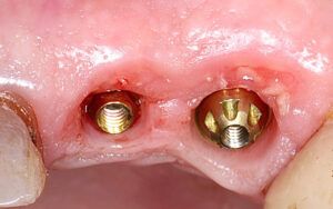
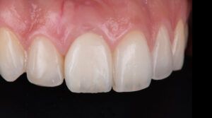
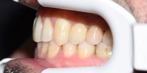
View of the healthy healed tissues View of the healthy healed tissues Definitive prosthesis in the mouth Definitive prosthesis in the mouth


Conclusion
According to the literature, from a biological point of view, preserving the socket in the placement of a post-extraction implant makes it possible to stabilise the marginal bone levels, making the survival of these implants comparable to those placed in fully healed crests.
The excellent response of the tissues in this case shows that the technique of immediate load and regeneration of the vestibular gap enables this soft tissue to be stabilised and the desired emergence profile modelled using the provisional prosthesis, transferring this outline to the definitive fully customised restoration.
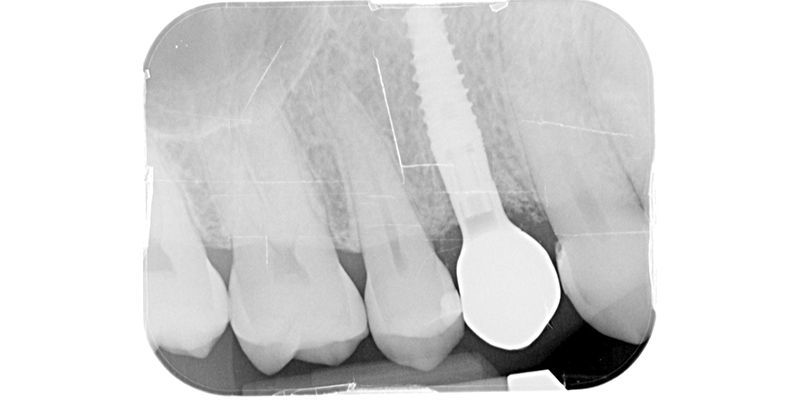
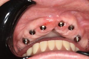
Image of the good condition of the tissues around the titanium base Details of the manufacture of the customised immediate provisional prosthesis Vestibular view of the provisional crown in the mouth, adapted to the gingival margin Occlusal view of the immediate-load provisional crown x-ray image of the provisional crown screw-mounted to the implant 
Design sequence of the full-volume crown Design sequence of the full-volume crown Design sequence of the full-volume crown Design sequence of the full-volume crown Design sequence of the full-volume crown Design sequence of the full-volume crown Images of the segmentation sequence of the design for the costumised definitive prosthesis Images of the segmentation sequence of the design for the costumised definitive prosthesis Images of the segmentation sequence of the design for the costumised definitive prosthesis Images of the segmentation sequence of the design for the costumised definitive prosthesis Images of the segmentation sequence of the design for the costumised definitive prosthesis Images of the segmentation sequence of the design for the costumised definitive prosthesis Details of the customised single restoration Details of the customised single restoration X-ray of the perfect fit of the structures and the outline of the interproximal tissue View of the healthy healed tissues View of the healthy healed tissues Definitive prosthesis in the mouth Definitive prosthesis in the mouth