Introduction
The geometry of the new Biomimetic Iceberg implant presents the ideal characteristics for designing restorations with implants and prosthetics that observe the biology of the hard and soft tissues, facilitating the aesthetics. The converging shape of the transmucosal part of the neck, with no angles or sharp edges, together with the new line of abutments, provides continuity to the neck of the implant increasing the generation of soft tissue, improving its preservation and achieving the adjustment profile established when planning the case.
Clinical case
We present a case of an 87-year-old patient who came to the clinic with pain in the right central incisor. The examination revealed an unstable crown. Further tests confirmed it was fractured and could not be repaired.
The treatment plan was decided, consisting of removing tooth 21 and placing an Avinent Biomimetic Iceberg immediate-load guided implant with regeneration of the vestibular gap, in conjunction with the placement of a customised provisional abutment with immediate load to stabilise the periimplant tissues during the osseointegration period.
A plan was made to place an Iceberg implant with a polished collar due to the thickness of the soft tissues that the patient presented. As this case affected an anterior aesthetic area, the quality of the tissues enabled us to distance the implant-prosthesis union from the bone, decreasing the risk of periimplant bone loss, and to model the tissue thanks to the tapered neck of the implant and the design of this prosthetic line.


Surgical and provisionalisation phase
An atraumatic extraction of tooth 21 was performed, respecting the alveolar walls, accompanied by a meticulous curettage of the socket to remove any pathological tissue.
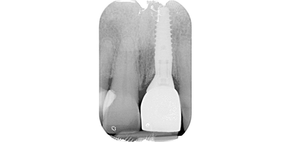
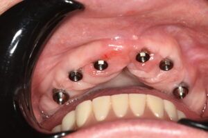
During the same surgical intervention, a Biomimetic Iceberg implant measuring 4 x 13 mm was inserted, guided by a splint resting on the teeth and designed with Implant Studio (3Shape) software.

Planning of the surgery guided by Implant Studio Details of the surgical spling Details of the surgical spling


Once the planned 3D position had been achieved, and the primary stability of the implant verified, a PEEK antirotation abutment was placed, on which the eggshell provisional was fitted to shape the desired emergence profile.
PEEK provisional screw-mounted on the implant showing the three-dimensional position PEEK provisional screw-mounted on the implant showing the three-dimensional position


The vestibular gap was filled with particulate biomaterial to avoid the reabsorption of the vestibular plate and finally the provisional tooth was screw-mounted at 35 Ncm, and it was confirmed that it remained out of the occlusion in maximum intercuspation and the excursive movement.
Preparation of the provisional with the PEEK abutment 
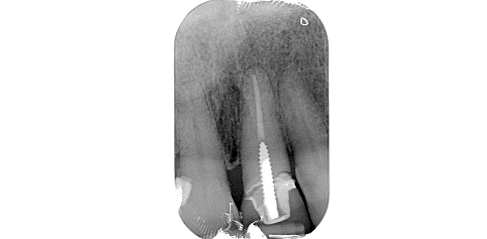
Preparation of the provisional with the PEEK abutment Finish of the provisional before placement Finish of the provisional before placement guide X-ray of the immediate provisional Details of the adjustment of the provisional restorarion in the mouth


Definitive prosthetic phase
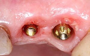
After a 3-month healing period the provisional restoration was unscrewed and the good periimplant health was verified. The tissues were scanned to transfer the information on the outline achieved during the provisionalisation period to the laboratory.
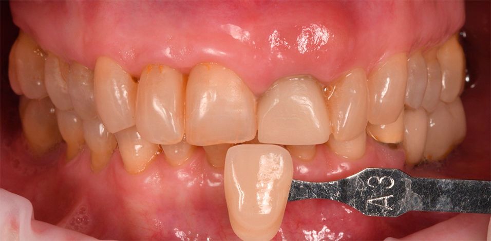
State of the tissues 3 months after implant placement and immediate provisionalisation State of the tissues 3 months after implant placement and immediate provisionalisation Digitally scanned abutment screw-mounted onto the implant and colour test


Scanned immediate-load provisional Scanned immediate-load provisional


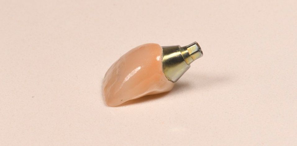
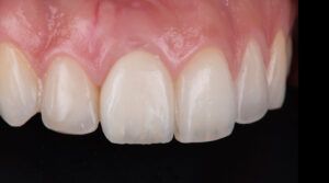
The definitive restoration on a customised titanium base where a zirconium crown with a ceramic coating was cemented for integration with the adjacent teeth.


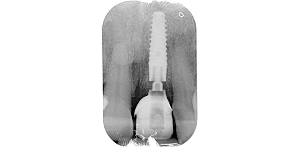
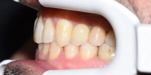
Health of healed periimplant tissues Health of healed periimplant tissues Image of the definitive restoration in the mouth


Conclusion
The good planning and execution of the case made it possible to obtain excellent clinical and aesthetic results, maintaining the gingival architecture with an immediate post-extraction implant and an immediate-load screw-mounted crown.
The design of the Iceberg implant enabled the biology of the tissues to be respected. A good thickness of keratinised gum and satisfactory 3D position of the implant led to highly pleasing results.
In this case the expectations of the patient were more than fulfilled; the symmetry of lower front teeth was improved, merely by replacing the fractured tooth.
Sequence of views of the final restorarion in the mouth Sequence of views of the final restorarion in the mouth Front view of the final restoration Front view of the final restoration